At a Glance
| Feature | Detail |
|---|---|
| Full name | Platelet-Rich Plasma Therapy |
| Mechanism | Concentrated autologous growth factors activate tissue repair |
| Most evidence | Knee OA, lateral epicondylitis, Achilles tendinopathy, androgenetic alopecia |
| Preparation method | Leukocyte-rich (LR-PRP) vs. leukocyte-poor (LP-PRP) |
| Platelet concentration | 3–8× baseline whole blood (optimum: ~1,000,000/µL) |
| Sessions typical | 1–3 injections, 4–6 weeks apart |
| Onset of effect | 4–8 weeks for musculoskeletal; 3–6 months for hair |
| Safety profile | Very high — autologous, no foreign proteins |
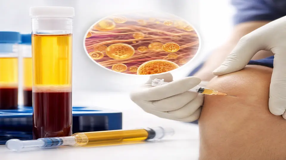
Platelet-rich plasma represents one of the more scientifically grounded regenerative interventions we use in integrative medicine. Unlike many interventions that rely on exogenous compounds, PRP harnesses your body’s own repair machinery — it simply concentrates and delivers it to where healing has stalled. Understanding when and how to deploy it is what separates good outcomes from disappointment.
The Biology: What Platelets Actually Do
Platelets are not simply clotting fragments. They are highly specialized cells carrying over 800 different proteins in their alpha granules, including growth factors that orchestrate every phase of tissue repair.
Upon activation — triggered by collagen exposure, thrombin, or the injection itself — platelets degranulate and release:
- Platelet-Derived Growth Factor (PDGF): Stimulates mesenchymal stem cell migration and proliferation; promotes angiogenesis
- Transforming Growth Factor-β (TGF-β): Regulates collagen synthesis and matrix remodeling; shifts macrophages toward anti-inflammatory M2 phenotype
- Vascular Endothelial Growth Factor (VEGF): Promotes new blood vessel formation into avascular tissue (tendons, cartilage)
- Epidermal Growth Factor (EGF): Stimulates epithelial and endothelial cell division
- Insulin-Like Growth Factor-1 (IGF-1): Promotes chondrocyte proliferation and type II collagen synthesis
- Fibroblast Growth Factor (FGF): Supports fibroblast activation and extracellular matrix deposition
Normal tissue relies on platelet concentrations of roughly 150,000–400,000 platelets/µL. A properly prepared PRP preparation achieves 1,000,000/µL or higher — a five- to sevenfold amplification that delivers these signals in concentrations tissue ordinarily never encounters.
Leukocyte Content: A Critical Variable
The presence or absence of white cells in the PRP preparation profoundly affects its biological behavior.
Leukocyte-rich PRP (LR-PRP) retains neutrophils and monocytes. It generates a more pro-inflammatory initial response and higher concentrations of proteolytic enzymes. This may be beneficial for chronically infected or fibrotic tissue (tendon, plantar fascia) where breaking down old matrix is necessary before rebuilding.
Leukocyte-poor PRP (LP-PRP) — also called pure PRP — removes most leukocytes. It delivers a cleaner growth factor signal with less initial inflammation. For intra-articular injections (knee, hip, shoulder), LP-PRP consistently outperforms LR-PRP because synovial tissue is sensitive to neutrophil-mediated catabolic enzymes.
This distinction is clinically meaningful. Meta-analyses that pool LR and LP studies produce muddier results than those that stratify by preparation type.
Where the Evidence Is Strongest
Knee Osteoarthritis
The most rigorously studied PRP application. A 2021 Cochrane review and multiple RCTs demonstrate that intra-articular LP-PRP provides superior pain relief and functional improvement compared to hyaluronic acid injections and corticosteroids at 6 and 12 months — with the additional advantage of no cartilage-degrading effects unlike steroids.
The RESTORE trial (2022, n=288) showed 78% of PRP-treated patients achieved minimal clinically important difference (MCID) in WOMAC scores at 12 months versus 51% in the saline group. Early Kellgren-Lawrence grades (I–III) respond best; Grade IV disease with bone-on-bone contact shows modest response.
Clinical protocol: LP-PRP, 5–7 mL per joint, 3 injections at 4-week intervals. Ultrasound guidance improves accuracy and outcomes.
Tendinopathies
Lateral epicondylitis (tennis elbow), Achilles tendinopathy, and patellar tendinopathy all have Level 1 evidence supporting PRP. The mechanistic rationale is compelling: tendons are hypovascular, and chronic tendinopathy is a failed healing response characterized by disorganized collagen, neovascularization, and absent inflammatory cells — precisely the state where delivered growth factors can restart repair.
LR-PRP appears superior to LP-PRP in tendon applications, consistent with the need for an initial inflammatory phase to recruit resident stem cells.
Clinical protocol: LR-PRP, 3–4 mL per site, 1–2 injections. Dry needling of the tendon immediately before injection improves growth factor uptake. Load management (eccentric protocol) must accompany injection for durable results.
Androgenetic Alopecia
Scalp PRP is now among the better-supported non-surgical treatments for pattern hair loss in both men and women. Growth factors from activated platelets — particularly PDGF, EGF, and VEGF — stimulate hair follicle stem cells, extend anagen phase, and promote miniaturized follicle recovery.
A 2019 systematic review of 19 controlled trials found significant improvements in hair count, hair thickness, and patient-reported satisfaction with serial PRP treatments. Results take 3–6 months to become visible; maintenance injections every 4–6 months preserve gains.
Clinical protocol: Intradermal scalp injections, 0.1 mL per site across the affected area, 3–4 sessions at monthly intervals.
Surgical Wound Augmentation and Chronic Wounds
PRP-infused matrices accelerate wound closure in diabetic foot ulcers, pressure injuries, and post-surgical sites with compromised healing. The evidence here is less from RCTs and more from clinical case series, but the biological rationale is robust.
In our practice, we routinely use PRP during complex wound management alongside systemic peptide support (BPC-157 and TB-500), as the combined local and systemic growth factor signaling appears synergistic based on mechanism and clinical observation.
Applications With Emerging Evidence
Intervertebral Disc Disease
Intradiscal PRP — injected into the nucleus pulposus under fluoroscopic guidance — has Phase II data supporting reduction in discogenic pain and disc height preservation. The nucleus pulposus is avascular and immunoprivileged; PRP growth factors can activate quiescent disc cells. Not yet standard of care, but a reasonable option for patients with MRI-confirmed disc degeneration and concordant pain who want to avoid fusion surgery.
Osteoarthritis of Other Joints
Intra-articular PRP for hip, shoulder, and ankle OA follows the same biological principles as knee. Evidence volume is lower, but mechanistic equivalence and clinical series support its use when conservative management has failed.
Post-Surgical Enhancement
Applied to the surgical site or injected perioperatively, PRP reduces time to healing, decreases infection risk (leukocytes in LR-PRP have direct antimicrobial activity), and improves tensile strength of repaired tissue. Used routinely in orthopedic surgery, maxillofacial reconstruction, and cardiac surgery.
How PRP Is Prepared: Process Matters
The preparation system significantly affects the final product. Not all “PRP” is equivalent:
-
Blood draw: Typically 15–60 mL of whole blood drawn under aseptic conditions, mixed with anticoagulant (sodium citrate preferred over EDTA — EDTA chelates calcium and impairs platelet activation)
-
First centrifugation (soft spin): ~300g for 5–10 minutes separates red cells from the “buffy coat” (platelets + leukocytes) and plasma above
-
Second centrifugation (hard spin): ~700g for 15 minutes concentrates platelets from the upper plasma fraction (produces LP-PRP) or from the buffy coat (produces LR-PRP)
-
Activation (optional): Calcium chloride or thrombin can pre-activate platelets before injection, converting PRP to a platelet-rich fibrin (PRF) gel. Activation enhances growth factor release but shortens the window for injection
-
Quality check: Platelet count of the final product should ideally be verified. Commercial systems like Arthrex ACP, Biomet GPS, or Harvest SmartPreP have validated preparation protocols with known yield characteristics.
Preparing for PRP Treatment
Two weeks before:
- Stop NSAIDs (ibuprofen, naproxen, aspirin) — they impair platelet function and blunt the inflammatory response necessary for healing
- Avoid corticosteroid injections for at least 4 weeks prior — steroids suppress platelet activity
- Maintain adequate hydration; hemoconcentration reduces PRP yield
Day of treatment:
- Eat normally — fasting is unnecessary
- Wear loose clothing over the injection site
- Arrange a ride home if heavy sedation is used (usually not required; most patients tolerate PRP with only local anesthetic)
After injection:
- Expect localized pain and swelling for 3–5 days — this is the intended inflammatory phase, not a complication
- Avoid NSAIDs for 2–4 weeks post-injection; acetaminophen is acceptable for pain management
- Ice can be used briefly for excessive swelling but may blunt the response — use sparingly
- For joint injections: relative rest for 48–72 hours, then graduated rehabilitation
- For tendon injections: guided physical therapy loading program starting at 2 weeks
Contraindications and Limitations
PRP’s autologous nature means systemic side effects are essentially absent — there are no allergic reactions, no disease transmission, no rejection. Local risks include infection (rare with aseptic technique), nerve injury (avoidable with image guidance), and temporary pain flare.
Absolute contraindications:
- Active systemic infection or bacteremia
- Platelet count below 105,000/µL (insufficient substrate)
- Platelet dysfunction syndromes
- Anticoagulation therapy that cannot be temporarily paused
Relative contraindications / reduced response:
- Heavy smokers (impaired platelet function and angiogenesis)
- Poorly controlled diabetes (dysfunctional platelets, poor healing environment)
- Chronic NSAID use not paused pre-treatment
- Advanced joint disease (Grade IV OA with absent cartilage)
Combining PRP with Peptide Therapy
In complex healing cases — post-surgical recovery, chronic tendinopathy, osteoarthritis with systemic inflammation — I combine local PRP injections with systemic peptide support. BPC-157 accelerates tissue repair through the same VEGF-angiogenesis pathway that PRP activates locally, creating complementary signaling. TB-500 upregulates actin polymerization critical for cell migration into the repair site. The combination addresses both the local growth factor environment (PRP) and systemic healing capacity (peptides).
For patients with systemic inflammation or autoimmune overlap driving tissue breakdown, I also evaluate whether immunomodulation — including low-dose naltrexone or targeted cytokine support — should precede PRP to optimize the healing environment.
Related Articles
- BPC-157: The Healing Peptide — Systemic healing support that complements local PRP injections
- TB-500 for Tendon and Tissue Repair — Peptide that enhances cell migration into PRP-treated sites
- Prolozone Therapy: Ozone-Enhanced Regenerative Injections — Ozone-based regenerative injection that can be combined with PRP
- BPC-157 vs. TB-500: Which Healing Peptide Is Right for You? — Comparing the two most used healing peptides alongside PRP
- What to Expect at Your First Integrative Medicine Consultation — How regenerative treatments fit into a comprehensive integrative plan
References
- Shen L, et al. “Leukocyte-poor versus leukocyte-rich PRP for knee osteoarthritis.” Am J Sports Med. 2021;49(8):2252-2261. PMID: 34110940
- Belk JW, et al. “Platelet-rich plasma versus hyaluronic acid for knee osteoarthritis: a systematic review and meta-analysis.” Am J Sports Med. 2021;49(1):249-260. PMID: 32644808
- Fitzpatrick J, et al. “The effectiveness of platelet-rich plasma injections in treating lower extremity tendinopathy.” Am J Sports Med. 2017;45(6):1453-1460. PMID: 27573323
- Alves R, Grimalt R. “A Review of Platelet-Rich Plasma: History, Biology, Mechanism of Action, and Classification.” Skin Appendage Disord. 2018;4(1):18-24. PMID: 29457008
- Everts P, et al. “Platelet-Rich Plasma: New Performance Understandings and Therapeutic Considerations in 2020.” Int J Mol Sci. 2020;21(20):7794. PMID: 33096812
- Kon E, et al. “Platelet-rich plasma: intra-articular knee injections produced favorable results on degenerative cartilage lesions.” Knee Surg Sports Traumatol Arthrosc. 2010;18(4):472-479. PMID: 19838676
- Moraes VY, et al. “Platelet-rich therapies for musculoskeletal soft tissue injuries.” Cochrane Database Syst Rev. 2014;4:CD010071. PMID: 24782016